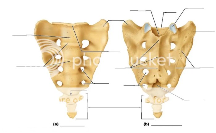
Label the structures of the sacrum and coccyx, an intricate exploration into the human body’s axial skeleton, unveils the fascinating anatomy of these interconnected bones. This journey delves into their unique shapes, articulations, and clinical significance, providing a comprehensive understanding of their essential roles in supporting and protecting the body.
Anatomical Overview of the Sacrum and Coccyx
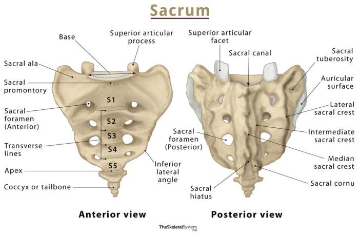
The sacrum and coccyx are located at the caudal end of the axial skeleton. The sacrum is a triangular bone formed by the fusion of five sacral vertebrae, while the coccyx is a small, triangular bone composed of four rudimentary vertebrae.
The sacrum is curved anteriorly and concave posteriorly, forming the posterior wall of the pelvic cavity. Its broad, superior base articulates with the fifth lumbar vertebra, and its apex points inferiorly.
The sacral vertebrae are numbered S1-S5 and have a general shape similar to the lumbar vertebrae. However, they are wider and have shorter, stouter transverse processes. The sacral canal is formed by the vertebral foramina of the sacral vertebrae and contains the sacral nerves and blood vessels.
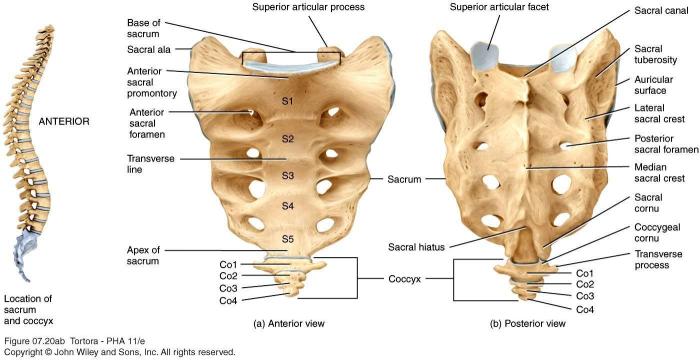
The coccyx is located inferior to the sacrum and is formed by the fusion of four rudimentary vertebrae, Co1-Co4. The coccygeal vertebrae are small and irregular in shape, and they gradually decrease in size from Co1 to Co4.
Sacral Structures
Sacral Alae
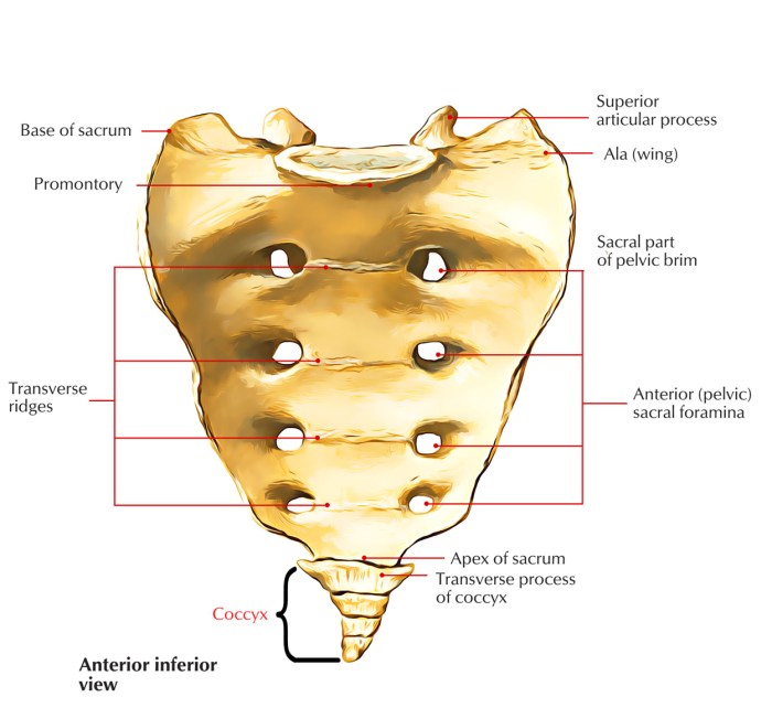
The sacral alae are the broad, lateral portions of the sacrum. They are formed by the fusion of the transverse processes of the sacral vertebrae and provide attachment points for muscles and ligaments.
Sacral Foramina, Label the structures of the sacrum and coccyx
The sacral foramina are located between the sacral vertebrae and transmit the sacral nerves and blood vessels.
Sacral Hiatus
The sacral hiatus is a large, oval opening located at the posterior aspect of the sacrum. It is formed by the incomplete fusion of the laminae of the sacral vertebrae and is covered by the sacrococcygeal ligament.
Coccygeal Structures: Label The Structures Of The Sacrum And Coccyx
Coccygeal Vertebrae
The coccygeal vertebrae are small, irregular bones that are fused together to form the coccyx. They are numbered Co1-Co4 and decrease in size from Co1 to Co4.
Coccygeal Cornua
The coccygeal cornua are small, lateral projections of the coccygeal vertebrae. They provide attachment points for muscles and ligaments.
Coccygeal Ligaments
The coccygeal ligaments are a group of ligaments that connect the coccygeal vertebrae and surrounding structures. They include the sacrococcygeal ligament, the intercoccygeal ligaments, and the coccygeal cornual ligaments.
Clinical Significance
Sacral Fractures
Sacral fractures can occur as a result of trauma or disease. They can be classified into three types: lateral compression fractures, vertical shear fractures, and burst fractures.
Lateral compression fractures are the most common type of sacral fracture. They are caused by a force that compresses the sacrum from the side, resulting in a fracture of the sacral alae.
Vertical shear fractures are caused by a force that shears the sacrum vertically, resulting in a fracture of the sacral body.
Burst fractures are caused by a force that bursts the sacrum open, resulting in a fracture of both the sacral alae and the sacral body.
Coccydynia
Coccydynia is a condition characterized by pain in the coccyx. It can be caused by trauma, childbirth, or prolonged sitting.
Symptoms of coccydynia include pain that is aggravated by sitting, standing, or walking; tenderness to the touch; and difficulty with bowel movements.
Pilonidal Cyst
A pilonidal cyst is a small, sac-like structure that forms in the natal cleft, the area between the buttocks.
Pilonidal cysts are caused by the accumulation of hair and debris in the natal cleft. They can become infected and painful.
Frequently Asked Questions
What is the clinical significance of sacral fractures?
Sacral fractures can result from high-energy trauma and may cause pain, neurological deficits, and instability of the pelvic ring, requiring prompt medical attention and specialized treatment.
What causes coccydynia?
Coccydynia, pain in the coccyx, can arise from trauma, childbirth, prolonged sitting, or underlying medical conditions, leading to discomfort and difficulty with sitting and bowel movements.
How are pilonidal cysts treated?
Treatment for pilonidal cysts typically involves surgical excision to remove the cyst and prevent recurrence. In some cases, antibiotics or other conservative measures may be employed.
